Hi everyone – You guys did great on Exam 3! The mean was 94% and everyone passed (yay!).
I’ve uploaded your scores for this exam in Canvas. I’ll figure out grades and post them shortly. If you have any questions about the exam, or the course, or your final grade, let me know.
It’s been so wonderful having you all in class. You’re so smart (obviously) but also kind, curious, compassionate, funny, and just a joy to be around. I’m so excited to have you guys again in Oral Histology, Embryology and Genetics, which starts in January.
Thanks for making this course fun (for me anyway!). I hope the rest of the semester goes really well for you. Take good care of yourselves!
This post contains specifics and logistics for our third exam in General Histology (DDS 6214).
Start and end time
The exam is scheduled for Tuesday, October 21, and you may take it any time between 12:00 am and 11:59 pm that day. Once you open the exam you have two hours to complete the exam. All submissions must be uploaded by 11:59 pm on Tuesday in order to receive a grade.
Password
The password for the exam is YourockD1s.
Classroom availability
Our classroom will be open and available from 1:00 – 2:55 pm on Thursday, if you want to take the exam at that time in our classroom. I’d suggest that if there are several people in the the room, please try to space yourself out well so there’s no way anyone can suggest the possibility of you looking at someone else’s computer. You can, of course, take the exam anywhere you want – so take it wherever you’re most comfortable.
Content
This exam has 39 points. Here’s the full breakdown by lecture for this exam:
All questions are multiple choice with one correct answer, and each question is worth one point. No photos or images.
One thing that I want to point out, just so they don’t trip you up: there are two questions that ask you “Which of the following DOES NOT ____” in the question stem. In general, I don’t like these questions (like: “all of the following EXCEPT”) because it’s easy to accidentally misread the question. But sometimes, it’s hard to come up with a reasonable question with reasonable answers without resorting to this method. So just a heads up.
Study resources
Please make sure to take a look at the following, which will help you focus on the material that is most important for you to know for this exam:
You will take this exam using Examplify installed on your PC/Macintosh laptop or desktop computer. If Examplify is currently installed, it may require an update and computer restart before the exam. If it is not installed, you should download and install the most up-to-date version of Examplify before the exam.
No scratch paper is allowed during the exam – but I have enabled the digital notepad feature within Examplify, so you can use that if that helps.
Also: this is not an open-book, open-note exam – and I trust that you will adhere to the honor code and not use any outside references.
Grading
After you take the exam, you will immediately receive your raw score. Final scores will be posted to your Canvas after I’ve reviewed the exam metrics.
Finally…
If you have any questions please feel free to email me (kkrafts@umn.edu) any time.
And if you need a little hype before you take the exam, here you go. You’re welcome.
I can’t help but draw analogies between Beyonce and female reproductive histology. I mean, it’s BEYONCE people. Here she is in all her glory at the 2017 Grammies:
She is adorned in a beautiful golden gown and her head is surrounded by a gorgeous radiating crown. Oh, AND she’s pregnant!
Beyonce is like a primary oocyte sitting in a Graafian follicle: her crown is the corona radiata, and her dress is made of gorgeous granulosa cells. At some point she no doubt sits on a little stool, which is the cumulus oophorus.
The metaphor continues! What happens during ovulation? Beyonce sheds her gown (the granulosa cells of the Graafian follicle) and leaves the ovary, making her way down that treacherous runway to the opening of the fallopian tube. So what’s left behind? Her gorgeous golden gown! That stays in the ovary, and becomes the corpus luteum (the “yellow body”).
I’m telling you, there’s even more here. If you watch the lecture recording, you’ll see I keep saying the Graafian follicle is juicy (maybe not the best description, but hey, that’s what it looks like to me!). And Bey is most certainly juicy – she straight up says it in Cozy!
This diagram shows the different maturational stages of ovarian follicles.
Here’s a quick summary of when you see these different stages:
At birth, all you have are primordial follicles (about a million in each ovary).
At puberty, you start “recruiting” groups of about 50 primordial follicles at the beginning of each menstrual cycle.
These primordial follicles start down the pathway of maturation, moving through the stages listed above (unilaminar primary, multilaminar primary, secondary, and Graafian follicles).
Only one of the group of 50 follicles will make it to the Graafian follicle stage (the other 49 die off along the way in a process called “attrition” or “atresia”).
At the time of ovulation, the Graafian follicle ruptures, releasing the germ cell inside, which makes its way to the fallopian tube and down into the uterus.
In addition, over a woman’s lifetime, the primordial follicles in each ovary start dying off on their own (independent of the menstrual cycles). Their population dwindles over time until the age of menopause, when there are none left. Then menstrual periods cease (there are no primordial follicles left to recruit!).
By the way, here’s what is inside each follicle (spoiler: it’s always a primary oocyte):
Primordial follicle: primary oocyte (arrested part way through meiosis I)
Unilaminar primordial follicle: primary oocyte (arrested part way through meiosis I)
Multilaminar primordial follicle: primary oocyte (arrested part way through meiosis I)
Secondary follicle: primary oocyte (arrested part way through meiosis I)
Graafian follicle: primary oocyte (arrested part way through meiosis I)
Wait, what?? What about the secondary oocyte and the ovum?
At ovulation, the primary oocyte is “released” from its maturational arrest, and it turns into a secondary oocyte. YAY! This secondary oocyte starts undergoing meiosis II, but is arrested part way through.
If no fertilization occurs, the secondary oocyte will never complete meiosis II, and will be shed during menstruation.
If fertilization occurs, the secondary oocyte completes meiosis II, turning into an ovum. FINALLY!!!
Here’s an interactive drawing I made that shows what happens to female germ cells over time. I think it’s kind of helpful to see the numbers (and stages) of the germ cells mapped out by age – it gives you a visual sense of what happens throughout life.
I can’t post the drawing on our website because of wordpress limitations – but I was able to post it on my Pathology Student website (so when you click on the link, you’ll wind up on that website).
Just a quick reminder: I am on vacation until the 25th, so for the next three weeks, we won’t have in-person class. I don’t like being away that long – it feels weird not seeing you guys at all – but I will do my best to keep checking in with you and I am always here if you have questions (just drop me an email).
Looking ahead a bit, this week is pretty simple – just two lectures (urinary and female reproductive systems) for you to watch at your convenience. The following week, we’ll have an asynchronous video lecture on the male reproductive system and an exam 3 review Kahoot for you to go through on your own.
I’ll keep posting stuff that I think might help you as you’re studying!
And now for something completely different, here are a few photos I took today and yesterday, in case you like seeing other people’s travel photos.
Right now, I’m in Sifnos, a small, sweet island not too far from Athens.
The view from where I’m staying, looking east.
So many cats here! This one almost got my fork.
Waiting outside my room for the sunrise this morning.
My two newest friends, so gentle and lovely, also just hanging out before sunrise.
Sunrise! If you look closely, you can see the outlines of Paros (in front) and Naxos.
Tomorrow’s asynchronous lecture video is about the urinary system. I thought I’d share this little self-study thing I put together on the tubular segments of nephron in case you find it helpful. It just asks you to name the segments, and recall what they do and what they look like. Totally optional – if it helps, great, if not, just ignore it!
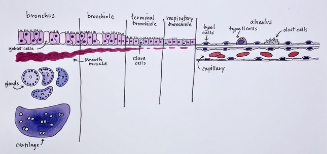
Here’s a drawing I made showing how the wall of the respiratory tree changes as you go from bronchi to alveoli. There are three versions, so you can use it fully labeled, partially labeled, or unlabeled, however it suits you best. Let me know if you find it useful – if so, I’ll make more 🙂
Q. I am slightly confused about muscularis externa. Is muscularis externa only present in places where muscularis mucosa is not? For example, the oral cavity, upper esophagus, and anal canal. Is it possible for them both to be present? And muscularis is not always present as a layer in the GI tract, correct? When present does it typically present in the same layer that the muscularis mucosa would be?
A. Those are great questions! As you noted, there are two “muscularis” layers in the GI tract: the muscularis mucosa and the muscularis externa.
The muscularis mucosa is a thin little layer of smooth muscle that is part of the mucosa (which includes epithelium, lamina propria, and muscularis mucosa). It innervates the inner layers of the mucosa, and it also shows you nicely where the mucosa ends, and the submucosa begins.
The muscularis externa is a thick layer of muscle that provides the main structural support all along the GI tract. It sits between the submucosa and the serosa/adventitia.
Basically the GI tract is just a tube with four concentric layers, like this:
From inside to outside, there’s mucosa (epithelium, lamina propria, and muscularis mucosa), submucosa, muscularis externa, and serosa/adventitia.
All levels of the GI tract have this exact structure, except for:
the mouth (which I’m not allowed to discuss with you lol)
the anus (which is only slightly different…its mucosa is comprised of just epithelium, with no lamina propria or muscularis mucosa).
I think that pretty much covers it. Having said all that, here are my direct answers (in blue)!
I am slightly confused about muscularis externa. Is muscularis externa only present in places where muscularis mucosa is not? For example, the oral cavity, upper esophagus, and anal canal. No. Both the muscularis mucosa and muscularis externa are present at every level of the GI tract except for mouth and anus (as mentioned above). The upper esophagus btw is not an exception – it has the same four-layered structure you see everywhere else. Is it possible for them both to be present? Yep! And muscularis is not always present as a layer in the GI tract, correct? No – both types of muscularis layers are pretty much always present. When present does it typically present in the same layer that the muscularis mucosa would be? No – the muscularis mucosa is always part of the mucosa (the innermost of the four layers), and the muscularis externa is always located between the submucosa and serosa/adventitia.